

Limb Atlas
Work in collaboration with Yong Wan and Chuck Hansen at The Scientific and Computing Institute, University of Utah.
Description of the methods for building the digital atlas of the developing mouse limb are now published in IEEE Computer Graphics and Applications' Special Issue - Biomedical Applications: From Data Capture to Modeling ![]() .
.
This is the link for the Tutorials for Making Anatomical Atlases.
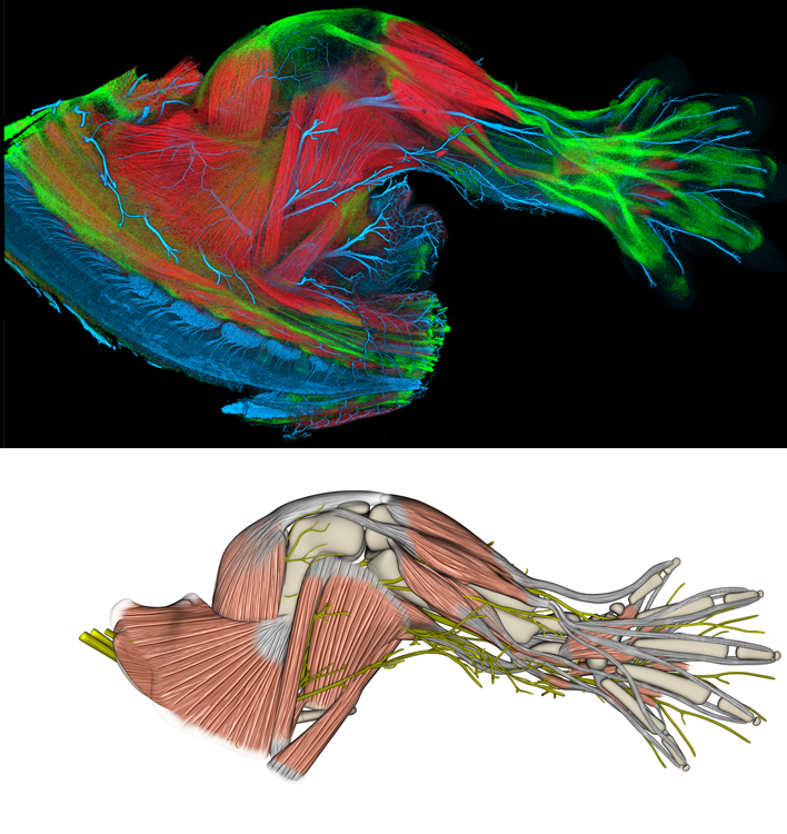
E14.5 mouse hind limb
Upper image is a confocal image of a limb in which muscle is labeled (in red) via immunofluorescence for myosin, tendons are labeled (in green) via ScxGFP transgene, and nerves are labeled (in blue) via immunofluorescence for neurofilament.
Lower image is the atlas view. In the upcoming atlas, the limb will be viewable in 3-dimensions. Also, all muscles, tendons, cartilage, and nerves will be labeled and individual components will be able to highlighted or removed, as desired.